

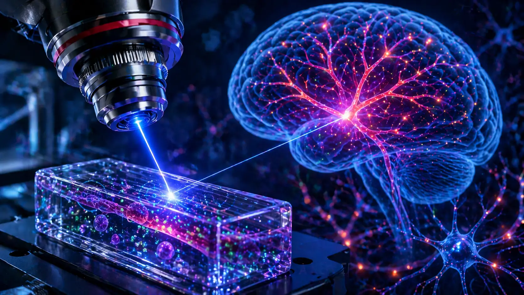
The MIT laser brain imaging breakthrough is redefining how scientists study the brain, offering a powerful new way to observe how drugs enter brain cells in real time. This cutting-edge discovery could significantly accelerate the development of treatments for neurological diseases by revealing whether medications actually reach their targets.
A Surprising Discovery in Laser Physics
Researchers at the Massachusetts Institute of Technology uncovered an unexpected phenomenon: chaotic laser light, which normally scatters unpredictably, can reorganize itself into a highly focused “pencil beam” under specific conditions.
This self-organizing beam challenges long-standing assumptions in optical physics. Instead of requiring complex adjustments, the system naturally produces a stable, precise beam when the laser is aligned perfectly and operated at high power levels.
25x Faster Brain Imaging
Using this new laser technique, scientists achieved 3D imaging of the blood-brain barrier up to 25 times faster than traditional methods—without sacrificing image quality.
The blood-brain barrier is a critical defense system that protects the brain but also makes drug delivery extremely difficult. Understanding how substances pass through it is essential for treating neurological disorders.
Watching Drugs Enter the Brain in Real Time
One of the most groundbreaking aspects of this innovation is its ability to track drug movement at the cellular level in real time.
Scientists can now:
- Observe how individual cells absorb drugs
- Measure how quickly medications penetrate brain tissue
- Identify whether treatments effectively reach their intended targets
This level of insight was previously impossible with conventional imaging techniques.
Why This Breakthrough Matters
This advancement could transform research into diseases like Alzheimer’s and ALS by solving one of the biggest challenges in medicine: verifying drug delivery to the brain. (ScienceDaily)
Key benefits include:
- Faster drug development cycles
- More accurate testing using human-based models
- Reduced reliance on animal studies
- Improved understanding of brain biology
Additionally, the method does not require fluorescent labeling, simplifying experiments and expanding its usability.
The Science Behind the “Pencil Beam”
To achieve this effect, researchers identified two essential conditions:
- Perfect alignment of the laser at a zero-degree angle
- High power levels that trigger interaction with the fiber material
When these factors combine, they create a balance that transforms disordered light into a clean, focused beam—eliminating common imaging issues like blur and distortion.
Future Potential and Applications
While still in the research stage, this technology has vast potential beyond brain imaging. Scientists are exploring applications such as:
- Imaging neurons and brain activity
- Studying complex biological tissues
- Advancing pharmaceutical testing
- Commercial biomedical imaging tools
As researchers continue refining the technique, it could become a standard tool in neuroscience and medical research.
Final Thoughts
The MIT laser brain imaging breakthrough represents a major leap forward in both physics and medicine. By enabling real-time visualization of how drugs interact with the brain, this innovation opens new doors for treating some of the most complex neurological diseases.
As the technology evolves, it could dramatically reshape how scientists understand the brain—and how we develop life-saving therapies.